Brief history

A HISTORY OF THE CHAIR
The Department of Human Anatomy was established in October 1945, when Kislovodsk Institute of Medicine was transferred to Chisinau. Based on its human resources Chisinau State Institute of Medicine (currently Nicolae Testemitanu State University of Medicine and Pharmacy) was founded.
1945-50
The Chair of Human Anatomy was established in October 1945, with the transfer to Chisinau of Kislovodsk Medical Institute, whose human resources laid the foundations of Chisinau State Institute of Medicine (CSIM), currently Nicolae Testemitanu State University of Medicine and Pharmacy.
The founder of the Chair and the first tenured specialist, who worked between 1945-1950, was Professor Emeritus A.P. Lavrentiev (1898-1958). Born in Vitebsk (Republic of Belarus), he came to Moldova from Kislovodsk (where he was the head of Department of Anatomy at the Institute of Medicine and an eminent specialist in the field of innervation of connective tissue formations, trained as anatomist at the famous school of anatomy of V.P. Vorobiov from Kharkiv (Ukraine), also the founder and head of the Department of Anatomy at the Medical Institute in Sverdlovsk (Ekaterinburg).
Among the first academic staff members of the Chair there were: assistant professors B.Z. Perlin, T.T. Koval, N.M. Volkova-Deineca (1945-1948), doctoral student P.I. Moskalenko (1945-1950), Associate Professor A.A. Shenfain (1946-1950) and assistant professor V.A. Tkaciuk (1947-1959).
Under the leadership of professor A. P. Lavrentiev: new dissection rooms and a laboratory were opened; the establishment of an anatomical museum was initiated, first exhibits being prepared; and an office for the study of radiological anatomy was set up.
Despite the extremely difficult conditions of the post-war period, the Chair was also concerned with scientific research in the field of vegetative innervation of the viscera, the implementation of macro-, meso- and microscopic methods for studying the morphology of tissues and organs, etc.
1950-59
In the years 1950-1951, the Chair was headed by Associate Professor V. Gh. Ukrainskii, who had a special interest in studying the synovial sheaths of the human hand tendons.
For three years (1951-1954) a considerable role in the development of the Chair was played by another disciple of scientist V.P. Vorobiov - university professor A.A. Otelin, under whose guidance: methylene blue staining of anatomic pieces according to V.P. Vorobiov’s method and silver impregnation technique, for the purpose of studying the innervation of the skin, the ileocecal region (B.Z. Perlin), were implemented and widely used; dissection methods have been unified; methodological guidelines for students have been developed; activities helping young teachers to achieve proficiency have been enhanced.
Starting with 1951, the teaching staff of the Chair was supplemented with young lecturers, CSIM graduates: assistant professor N. V. Cherdivarenco, G. V. Kucerenko-Vincenko (1951-2018); doctoral students V. Jiţa and A. Popa (1952-1997); assistant professor A. L. Kolesnik (1952-1957), L. A. Luneova (1952-1954), M. T. Selin (1952-1954), Iu. Takes. Muhin (1952-1953), and since 1954 - doctoral student N. Cereş (since 1957 - assistant professor).
In 1953, the Moldovan branch of the All-Union Scientific Association of Anatomists, Histologists and Embryologists (since 2013 - Public Scientific Association of Morphology of the Republic of Moldova) was created. It was led initially by Professor B.Z. Perlin and then by university professors V. Jita, M. Ştefaneţ, V. Nacu and I. Catereniuc.
Between 1954-1956, the Chair was headed by Professor Emeritus Valentina F. Parfentieva, representative of the School of Operative Surgery and Topographic Anatomy, founded by Professor V.N. Shevkunenko from Leningrad / St. Petersburg, a specialist in the angioarchitecture of endocrine glands and the viscera. Scientific research initiated during this period focused on surgical anatomy of blood vessels.
An important period in the scientific-didactic activity of the Chair begins with the appointment at its helm of university professor Vasilii V. Kuprianov (1956-1959), later, academician of the AMS of the USSR, Laureate of the State Prize of the USSR, President of the Society of Anatomists , Histologists and Embryologists of the USSR, editor-in-chief of the journal Archive of Anatomy, Histology and Embryology, a renowned specialist in the field of microcirculation.
Professor V. V. Kuprianov promoted new scientific directions both in the field of innervation of blood vessels and connective tissue structures and in the transcapillary and juxtacapillary circulation at microcirculatory level. In 1977, academician V.V. Kuprianov received the State Prize for a cycle of works on microcirculation issues. On the 90th anniversary of the Alma Mater, the scientist was awarded the title of Doctor Honoris Causa of Nicolae Testemitanu University for his special merits.
Representative of V. N. Tonkov’s anatomy school of Leningrad (St. Petersburg) and disciple of scientist B. A. Dolgo-Saburov, V. V. Kuprianov contributed significantly to the activation of scientific research and modernization of the discipline study. His scientific ideas were further developed in a series of PhD theses in medical sciences by V. Jita (1958), A. Popa (1958), N. Ceres (1961), GV Kucerenko-Vincenco (1961), N.V. Cherdivarenco (1961) and several Doctor Habilitated dissertations by B.Z. Perlin (1967), V. A. Tkaciuk (1970), V.Jiţa (1971), N. V. Cherdivarenco (1977), V. Andrieş (1989).
Between 1959-87, Associate Professor Boris Z. Perlin, Doctor Habilitated in Medical Sciences, university professor, MSSR Scientist Emeritus, fronted the Chair. Under his guidance, the fruitful activity of the Human Anatomy Chair continued.
During that period, multilateral research was conducted in order to establish morphological regularities as regards the peripheral innervation of connective tissue structures and blood vessels.
B.Z. Perlin used for the first time the macro-microscopic method of fine dissection of total anatomical parts, selectively stained with Schiff's reagent, to study the peripheral nervous system and implemented in scientific activity other methods of highlighting nerve structures (impregnation, histochemistry, luminescent).
Under the leadership of university professor B.Z. Perlin, 14 PhD theses and one Doctor Habilitated dissertation in medical sciences were defended, and over 100 scientific papers, 2 collections, 2 monographs, a dissection guide, a guide in the field of angioneurology and others were published.
For special merits, university professor B.Z. Perlin was decorated with the medal For Valiant Labour in the Great Patriotic War 1941–1945, the Eminent Health Care Employee badge, honorary diplomas, and the Veteran of Labor medal.
With the organization of the faculties of Dentistry, Preventive Medicine, Pharmacy and the increase in the number of enrolled students, the process of selecting and training young teachers has expanded.
Throughout that time, N. Fruntaşu (1961-1964), IV Kuznetsova (1960-1987), A. Nastas (1963-1975), V. Covaliu (1963-1977, 2002-2015), V. Andrieş (1964-2011), M. Casian (1964-1966), M. Arventeva (1964-1966), T. Lupaşcu (1965), M.Ştefaneţ (1965), V. Corduneanu-Covaliu ( 1965-1995, 2002-2017), D. Didilica-Stratilă (1965-2000), G. Marin-Hâncu (1966), EG Sadovaia (1966-1968), I. Bostan (1966-1978), I.Guriţencu ( 1967-1976), E. Gherghelegiu-Poburnaia (1967), Gh. Nicolau (1969-1973), E. Beşliu-Lopotencu (1970), D. Batâr (1971), T.M. Titova (1974-2017), I. Catereniuc (1980), L. Gurieva-Mazilu (1982-1986), O.V. Belic (1987) etc. were added to the staff list of the chair.
Back then, the professional training of young lecturers occurred through secondary clinical education, doctoral and postdoctoral studies and refresher courses held at various medical institutes in Moscow, St. Petersburg, Kiev, etc.
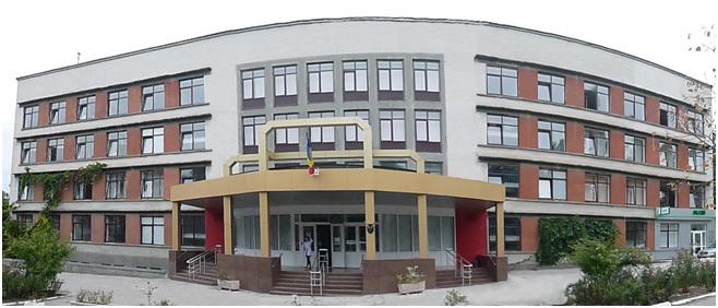
In 1965, the Chair moved to the current Morphological Block. The new building offered optimal conditions for the study of the discipline and for the arrangement of the anatomical museum, which was considerably expanded later, becoming very popular.
The images of impeccable quality of anatomical pieces, made by academic staff members (prof. B. Perlin, V. Andrieş, N. Cherdivarenco, M. Ştefaneţ, I. Catereniuc, Associate Professor G. Vincenco, E. Gherghelegiu- Poburnaia, T. Lupaşcu, E. Lopotencu, assistant D. Stratilă, Z. Zorina, etc.) following scientific investigations and exhibited in the museum halls, were used to illustrate many specialized editions, published in the country and abroad, including the famous Atlas of Human Anatomy (Sinelnikov R. D., Sinelnikov Ia.R. Atlas of Human Anatomy. Vol.4, Moscow, 1989 and The Vegetative Nervous System Atlas by Lobko P.I., Meliman E.P., Denisov S.D., Pivchenko P.G., Minsk, 1988)
The scientific research work carried out during that period resulted in 5 Doctor Habilitated theses by B. Z. Perlin (1967); V. Jita (1971); N. V. Cherdivarenco (1977); V. Andrieş (1988); M. Ştefaneţ (1998) and over 30 PhD theses developed by N. Fruntaşu (1964); IV Kuznetsova (1965), A. Nastas (1969), M. Chiorescu (1970), V. Andrieş (1970), V. Covaliu (1971), T. Lupaşcu (1972), M. Ştefaneţ (1972), Gh. Nicolau (1973), V. Voloh (1973), D. Batâr (1980), E. Beşliu (1988), E. Poburnaia (1993), I. Catereniuc (1998) etc.
During the Soviet period, two anatomy textbooks were translated into Romanian (Textbook of normal human anatomy by Lysionkov N.K., Bushkovich V.I., Prives M.G.; translated by V. Jița, A. Popa, M. Casian, T. Lupașcu and Human anatomy by M.R. Sapin in 2 volumes, translators M. Ștefaneț, T. Lupașcu, D. Batâr, both edited by D. Stahi.
1990-97
From 1988 to 1990, the Chair is headed by Associate Professor Mihail Ştefaneţ, currently Doctor Habilitated in Medical Sciences, university professor, Scientist Emeritus, specialist in periosteum innervation and issues related to the morphology of the funiculo-testicular complex.
M. Ştefaneţ made a special effort to reorganize and furnish the anatomical museum (including the opening of the Anatomy of the Child room) and to open a computer room, necessary for the implementation of virtual anatomy programs.
Under the leadership of M. Ștefaneț, 2 PhD theses and 3 Doctor Habilitated theses in medical sciences were defended.
It is worth mentioning the three volumes of Human Anatomy textbook (author- university professor M. Ştefaneţ), published and re-printed in recent years.
For the next six years (1991-1997), the Chair was headed by Doctor Habilitated in Medical Sciences, university Professor Emeritus Vasile Andrieş.
His experimental studies show eloquently the presence of multiple connections between the nerve plexuses of the viscera in the thoracic and abdominal cavities, connections that ensure collateral innervation of the lungs and largely explain the nature of repercussions on the lungs after abdominal organs surgery.
Prof. univ. V. Andrieş has published over 240 scientific papers (monographs, textbooks, methodological-didactic papers, articles and theses) in the country and abroad.
Other works of significance are Human Anatomy and Head and Neck Anatomy textbooks developed jointly with well-known representatives of specialized universities of Oradea and Timisoara (Romania). Under his leadership 3 PhD theses have been completed.
During the mentioned period, the following young assistants started their activity at the department - M. Pleşca (1990-1999), V. Lupu (1990-1993), L. Spataru (1990-1992), A. Vîlcu (1991-1994), G. Certan (1991), Z. Zorina (1991), S. Cheptănaru (1992-2006), A. Covalciuc (1992-1995), A. Antoci-Babuci (1993), N. Lozovan (1993-1995), V. Supciuc (1994-2007), T. Carajia-Botnari (1993), A. Bendelic (1996), A. Ioniţa (1995-2005), T.V. Hacina (1996), L. Globa (1997).
In 1997, in order to streamline the teaching process at the faculties of General Medicine, Pediatrics, Dentistry, Pharmacy and Preventive Medicine, two subdivisions were set up within the Department: one for the faculties of Pharmacy, Dentistry and Preventive Medicine, led by university professor Vasile Andrieș, and the other for the faculties of General Medicine and Pediatrics, headed by Mihail Ştefaneț (since 1998 Doctor Habilitated in Medical Sciences and since 2001 – university professor, Professor Emeritus of public education).
From 1997 to 2007, Human anatomy Chairs no. 1 and no. 2 operated separately, having the same headquarters and material-didactic base.
Between 1997-2007, the analytical program of the discipline was finalized, with emphasis on the study of living anatomy and the applicative aspect of the studied structures, methodological indications on living anatomy and questionnaires were elaborated, the forms of knowledge assessment were modernized, sets of control tests were developed in Romanian, Russian and English, and conditions for groups with teaching in French and English were created.
Much emphasis was placed on the professional training of young teachers, who were educated within the Chair, under the guidance of experienced professors, as well as on the refresher training at the anatomy departments of medical universities in Romania - Iasi, Cluj-Napoca, Bucharest, Targu -Mureş, Timişoara, Sibiu etc.
Subsequently, the Chair has been staffed by new graduates of Alma Mater: S. Brenişter (2007), R. Angheliu (2009), M. Taşnic (2011), I. Stupac (2011-2016).
Years 2013-20
In 2013, Ilia Catereniuc, Doctor Habilitated in Medical Sciences, university professor, has been elected as Chief of Chair by competition.
The objectives set before the Department's staff during this period are the implementation of the most optimal and progressive, in terms of immediate application, methods of training and assessment of students' knowledge, as well as the reasonable selection of scientific information included in the study process, aiming at the continuous improvement of the quality of medical education and students’ knowledge acquisition which will allow them to carry out their professional activity at the highest level.
The programs, curricula and questionnaires for the current and intermediate assessment of students' knowledge at all faculties were revised and updated (Medicine no. 1, Public Health specialties, Optometry and General Health Care, Medicine no. 2, Dentistry and Pharmacy)
It was imperative that applied anatomy be thoroughly studied given the advanced imaging diagnostic methods. Surface anatomy, projection of organs onto the body surface, study of cross- sectional anatomy, general principles of anatomical formations study through modern clinical and paraclinical techniques, live anatomy represent an indispensable component of the study of human body morphology, which guides the student towards a deeper perception of the links between form and function, contributing to the replacement of the static concept with a dynamic one in the acquisition of anatomical data.
Some outdated theoretical notions concerning the structure of anatomical formations, which are not essential in the professional training of doctors have been excluded from the curriculum, the requirements on the applicative importance of a series of anatomical structures with a minor practical impact were revised and the programs were brought into line with contemporary requirements.
Changes have been included in the study programs for the Faculties of Medicine, Dentistry and Pharmacy taking into account the faculty profile and the aspect of applying the knowledge. For this purpose, at the Faculty of Pharmacy and at the Faculty of Dentistry, a new curriculum has been developed and the study plans have been altered and implemented in the academic year 2014-2015.
To ensure the integration of virtual training programs in the discipline curriculum, the Chair has reorganized the computer room, which is equipped now with 16 high-performance computers, used both for teaching purposes (showing 3D video films for virtual training, practically for all discipline sections) and for student testing.
The chair’s collection of tests and situation problems has been developed for the first time in the country’s medical didactic literature, as all the provided correct answers to them are accompanied by state-of-the-art scientific arguments. The idea of this work belongs to its author - Associate Professor T. Lupașcu. This collection is intended for the individual work of students, but can also be used by practitioners and other specialists in the field to verify and update their anatomical knowledge.
In the context of studying the discipline of human anatomy in two semesters and due to the need to accelerate the extracurricular activity of students, for the development of teamwork and self-training skills, for the assessment and self-assessment of students, the Chair has a room with anatomical preparations on display (corpses with muscles, dissected vessels and nerves, organ systems and separate organs, molds, etc.), which is used during both practical classes according to the schedule and independent work of students.
Corpses with dissected muscles, vessels and nerves, preserved by the technique of plastination, polymer embalming with viscous substances, have been purchased. Plastinated anatomical preparations have a number of important qualities: they are non-toxic, odorless, mantain their natural shape and color, are demonstrative, can be used for a long time, do not require additional processing and storage capacity, are durable, body parts and the limb segments are mobile, their use is financially convenient.
Under the project of opening the University Center for Simulation in Medical Training, in order to ensure the didactic process, an impressive number of molds of different organs has been procured.
These progressive directions in the organization of the didactic process are elucidated in the methodical-didactic works published in the last years by the academic staff members: textbooks, guides for practical works on Human Anatomy (sem. I-II), collections of courses, collection of schemes for human anatomy, collections of tests and situational problems in human anatomy, etc., which are intended to facilitate the preparation of students for practical work, to implement contemporary training methodologies and to verify and deepen their knowledge in the field of anatomy.
It is worth mentioning the special methodical indication for self-training and self-assessment, developed by the staff of the Chair in order to capitalize on individual student work, coordinate and unify the educational process, which is published at the beginning of each semester (Practical works on Human Anatomy / Workbook in Human Anatomy / Практические занятия по анатомии человека; Ghid pentru autoinstruire / Guide for self-studying / Пособие по самоподготовке. I-II Vol.).
As regards independent work, conditions are created for the involvement of students in the dissection of muscles, vessels and nerves, organs, etc. during practical classes and at the Scientific Circle, led in different periods of time by assistant V. A Tkaciuk, Associate Professor T. A. Iastrebova, A. Popa, T. M. Titova, E. Lopotencu, V. T. Supciuc, University professor T. Hacina and, currently, by University professor O. Belic.
Here young researchers make the first attempts to apply the methods of anatomical investigation, study the variants and anomalies of development of organs, blood vessels and nerves, vascularization and innervation of anatomical formations, live anatomy, etc., gain the necessary experience in preparing reports and scientific studies. , actively participates in the proceedings of congresses, conferences and scientific symposia in the country and abroad, publishes scientific papers.
Many of the anatomical pieces made by the students are exhibited in the museum of the Department.
The development of the Museum of Human Anatomy remains a priority. It has one of the most valuable and imposing collections of unique anatomical pieces (about 2000 exhibits), displayed in 5 spacious rooms (Locomotor System; Viscera; Central Nervous System; Vessels and Nerves; Anatomy of the Child), being one of the few this kind in Europe, highly appreciated by many specialists from abroad, who visited it.
At the initiative of Prof. I. Catereniuc, a meticulous work of organizing and enriching the museum with new exhibits is carried out. The Electronic Catalog of Anatomical Exhibits of the Museum, placed on the Department's WEB page, is currently being worked on.
In recent years, new Alma Mater graduates joined the academic staff of the Department: Pasha D., (2013) Stegarescu Ion (2014), Vrabii Vitalie (2015), Gînju Nadia (2016), Ursu Alexandru (2017) , Negarî Nadejda (2017).
From its foundation until now, the Department scientific topics are primarily concerned with the study of the innervation of periosteum of bones, capsules and ligaments of various joints, lepto- and pachymeninges, the innervation of connective tissue formations and main blood vessels, and of internal organs in normal and pathological condition; In laboratory animal research, the influence of metered physical loading, hyper- and hypokinesia and hyperbaric oxygen therapy was studied.
From 1997 on, the scientific activity at the Department has been continually gaining impetus - 4 Doctor Habilitated theses (M. Ştefaneţ, 1998, I. Catereniuc, 2007, O. Belic, 2017, T. Hacina, 2017) and 6 PhD theses in medical sciences (I. Catereniuc, 1998, V. Supciuc, 2000, G. Certan, 2003, V. Focşa, 2003, T. Hacina, 2004, O. Belic, 2005) have been defended.
Assistant professors A. Babuci, A. Bendelic, D. Pașa, M. Tașnic, Z. Zorina, R. Angheliu V. Vrabii, I. Stegarescu, L. Globa, N. Negarî are currently studying for PhD degree.
In order to create better conditions for conducting scientific research, a morphological laboratory endowed with appropriate equipment has been set up.
Over the years, the scientific interests of the Department’s academic staff have been expanding according to the new requirements in the area.
The main direction of scientific research has gradually taken shape: Innervation of connective tissue formations in normal and pathological state; Morphology of para-, perivisceral and perivascular elements; The specifics of the ligament system of the internal organs and their biomechanical properties, the morphofunctional peculiarities of different organs and systems in the critical periods of postnatal development; Individual anatomical variability, correlations and morphoclinical features of blood vessels and peripheral nerves.
At present, the Department has established fruitful didactic-scientific cooperation relations with colleagues from similar departments of medical universities in many CIS countries and Europe (Minsk, Grodno, Vitebsk, Chernivtsi, Kiev, Ivano-Francovsk, Bucharest, Iasi, Constanta, Cluj-Napoca, Timisoara, Moscow, Smolensk, Varna, Tbilisi etc).
The auxiliary staff (trainers, laboratory assistants, senior laboratory assistants), whose help and support is felt by each member of the academic staff, plays a significant role in the daily life of the Chair.
Among the auxiliary staff members who made a visible contribution to the development of the didactic-scientific activity of the Chair, the following specialists should be mentioned: senior anatomical preparator I.D. Popazov (1946-1976), museum chief, senior laboratory assistant A.V. Leşcenko (1959-1977), senior laboratory assistant L.P. Stasieva, senior laboratory assistant V. Ceban, preparator / painter M. Lupu, laboratory assistant M.A. Savina, preparator A. Ababieva, senior laboratory assistant Cleopatra V. Ciornaia-Leonidova, senior laboratory assistant V. Bondarenco, senior laboratory assistant V. Şevciuc, preparator V.V. Sobeţkaia (1964-1985), senior laboratory assistant E.M. Koblik-Zelţer (1968-1989), laboratory assistant L.N. Samoşina (1967-1989), senior laboratory assistant M.L. Serbova (1972), senior laboratory assistant N. Bujoreanu (1972), senior laboratory assistant D. Pâslari (1973-1995), museum chief, senior laboratory assistant J.I. Pavlenko (since 1977), laboratory assistant A. Popa (1987-1990), laboratory assistant T. Romanenko (1991-1995), laboratory assistant R. Frumusachi (1991-1995), preparator VN Burlacic (1991-1995) , preparator P. Stratilă (1993-1996), laboratory assistant L. Balan (from 19 93), preparator P. Stețenco (since 1997), laboratory assistant L. Poiana (1996-2003), laboratory assistant G. I. Drotieva (1999-2001), preparators E. Popa (since 2017) and Z. Timotin (since 2017).
