
Museum of Human Anatomy
Room 1.1. LOCOMOTOR SYSTEM (exhibits).pdf
Room 1.2. LOCOMOTOR SYSTEM (exhibits).pdf
Room 2.1. VISCERA (exhibits).pdf
Room 2.2. VISCERA (exhibits).pdf
Room 3. CENTRAL NERVOUS SYSTEM (exhibits).pdf
Room 4.1. VESSELS and NERVES (exhibits).pdf
Room 4.2. VESSELS and NERVES (exhibits).pdf
Room 5.1. ANATOMY Of CHILD. TERATOLOGY (exhibits).pdf
Room 5.2. ANATOMY OF CHILD. TERATOLOGY (exhibits).pdf
THE MUSEUM OF ANATOMY
from the State University of Medicine and Pharmacy
"Nicolae Testemitanu" - a remarkable collection of anatomical pieces
The Anatomy Museum within the Department of Human Anatomy of the State University of Medicine and Pharmacy "Nicolae Testemitanu", one of the richest in Europe and the post-Soviet space, represents more than just a presentation of exhibits, it is a unique collection , internationally, thanks to the number and quality of the anatomical parts it contains. He is a real pride and, at the same time, a priority for the University and, not least, for the Department of Human Anatomy.
We are talking about a certain type of museum - the Museum of Anatomy - which contains numerous anatomical pieces of extraordinary quality, made over time by various processes (fine anatomical dissection, corrosion, mummification, plastination, etc.) by the scientific-teaching staff who they have worked over the years and currently work at the Department of Human Anatomy of the University, contributing both to its renovation and enrichment with new exhibits, as well as to its implementation in the process of educating students.
The idea of organizing an Anatomical Museum appeared in the post-war period in the first years after the establishment of the Department of Human Anatomy (October 1945), with the transfer of the State Institute of Kislovodsk to Chisinau, based on the human potential of which the State Institute was founded of Medicine from Chisinau, currently USMF Nicolae Testemitanu, an idea that was put into practice by the first holder of the chair Alexei P. Lavrentiev (1945-1950) and the assistant, then, Boris Z. Perlin (future chair of the chair - 1959- 1987).
The first and oldest exhibits in the museum date back to that period and refer to the locomotor apparatus (bones, spine joints, upper limb muscles, etc.).
During the period 1965-1971 the museum was reorganized, enriched and completed with an imposing number of new exhibits. Currently, the museum has one of the most valuable collections of unique anatomical pieces, which are complemented daily with new exhibits, being one of the few of its kind, highly appreciated by specialists in the field in the country and abroad.
It should be mentioned that the museum is the result of the activity of both the holders, who have been at the helm of the Chair for different periods of time: Alexei P. Lavrentiev (1945-1950), V. Gh. Ukrainski (1950-1951), AA Otelin (1951- 1954), Valentina F. Parfentieva (1954-1956), Vasilii V. Kuprianov (1956-1959), Boris Z. Perlin (1959-1987), Mihail I. Stefanţ (1988-1990) (1998-2013), Vasile N Andrieş (1991-1997), Ilia M. Catereniuc (2013 - present), as well as anatomy teachers - Natalia V. Cherdivarenco, Galina V. Vincenco, Victor T. Jița, Alexei V. Popa, Tatiana A. Iastrebova, Ilia A. Gurițenco, Anatolie N. Nastas, Teodor I. Lupașcu, Tamara V. Titova, Dumitru Gh. Batîr, Eugenia S. Lopotencu, Emilia V. Poburnaia, Olga V. Belic, Tamara V. Hacina, Zinovia A. Zorina, Tatiana C. Botnari and the workers - NV Lescenco, Janna I. Pavlenco.
For more than seven decades, the pieces of collection have supported the transfer to a new building (1965), have passed through a series of earthquakes, giving the possibility and creating optimal conditions for students for the theoretical and practical study of anatomy.
The young students also participate in the arrangement of the rooms. Some of them, members of the Student Scientific Circle, were and are involved in the dissection process - the anatomical pieces made by the young doctors complement harmoniously the impressive collection of anatomical pieces exhibited in the museum.
The museum is extremely rich in teaching material. The students first learn in the dissection rooms (the room for demonstration and study of the anatomical pieces arranged at the Department), and for the demonstrative supplementation of the information regarding the anatomical formations - they study here.
The Museum of Anatomy within the USMF "Nicolae Testemitanu" besides the main purpose - didactic and scientific, represents also a starting point in understanding the medical phenomenon for those who are curious and passionate about the inner beauty of the human body. Thanks to these exhibits the mysteries of human creation can be understood.
The museum enjoys great popularity being visited by numerous students and teachers of the high schools, by the students of colleges and universities in the country and by numerous delegations from abroad, by representatives of different public organizations etc.
One fact we are proud of is that, during Open Doors Day, the museum is presented to future medical students, for many of whom it is an impulse to follow this professional path.
Moreover, the museum plays a special role in popularizing knowledge about Man, regarding morphofunctional particularities at different stages of pre- and postnatal ontogeny, individual anatomical variability and the influence of different environmental factors on the structure and activity of organs and organ systems. and so on
That said, in addition to the cognitive value of such anatomical pieces, the museum is also of real educational use.
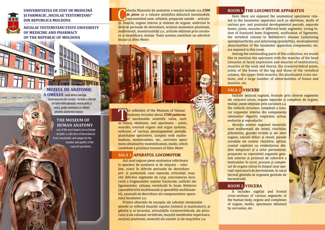
The collection of the Museum of Anatomy includes about 2000 pieces with an invaluable scientific-didactic value, representing bones, skeletons, wet preparations - joints, muscles, internal organs and organ systems, embryos in different periods of development, anatomical sections, plastinations, malformations, monstrosities etc., kept in solution of formol, mummified parts and molds.
The unique anatomical pieces, exhibited in 5 spacious rooms within the Department of Human Anatomy (the locomotor apparatus; Viscere; Central nervous system; Vessels and nerves; Child's anatomy), constitute a true treasure of the Alma Mater.
Room 1. The locomotor apparatus
In this room you can admire a rich collection of anatomical pieces related to the support and movement apparatus - skeletons, skulls in different periods of pre- and postnatal development and separate bones, joints, muscles of different body segments, incorrect concretion of fractured bone fragments , ossification of the ligaments, spine in Behterev's disease (ankylosing spondylarthritis and ankylosing spondylitis), developmental abnormalities of the locomotor system components, etc.
Among the exceptional pieces of this compartment of the collection we mention the piece that reflects the muscles of the head (mimics / masticators), of the neck and the chest, the craniovertebral joints, the anatomical sections plated through the joints of the foot, an imposing number of anomalies of the bones and muscles etc.
Here we find exhibits and the first exhibits, which laid the foundation for the future museum - the joints of the spine, the muscles of the upper limb, as well as several similar pieces made in subsequent periods.
Also worth mentioning are the anatomical pieces made over the years by students, members of the Scientific Circle, who occupy a special place in two showcases.
*
Room 2. Viscera
The anatomical pieces on display in this room include sagittal, frontal sections, etc. through various segments of the human body, separate and complex organs, molds, corrosion parts, etc. They reflect the complex structure of all the internal organs of the digestive, respiratory, urinary, endocrine and reproductive systems.
The attraction of this segment of the museum collection are malformations of the heart, kidneys, lungs, thyroid gland and other organs, gall and kidney stones, corrosion parts of the vascular systems of the kidneys, spleen, etc.
Needs attention and the exposed - the skull of the child with the highlighting of the temporary and the mature teeth, with the highlighting of the permanent ones, the preparations that reflect the external genital organs and the process of descending the testicles in the scrotum and the complex of organs extracted during a herniotomy repair operation, in the hernial sac, the genital organs of the hermaphrodite.
*
Room 3. Central nervous system
The exhibits in this room reflect in different aspects all the structures within the central nervous system - sections in different projections of the encephalon (rhombencephal with its components, structure of the mesencephalon and of the proencephalon with its constituent elements, cerebral ventricles) and of the spinal cord, including the cerebral and spinal meninges. with their derivatives.
Among the pieces in this compartment of the collection are the sections through the cerebral and cerebellar hemispheres, colored by the precipitation of cobalt sulphide to highlight the relationship between the white and the gray matter, the spinal cord of a child over its entire length, the open cerebral ventricles, etc.
*
Room 4. Vessels and nerves
It is of great historical value, because it houses a very diverse collection of anatomical pieces related to the innervation and vascularization of the various organs, proof of the continuity of the research in the field of anatomy, preparations made by the scientific-didactic staff of the Chair during the research activities regarding the elaboration of theses. doctor / doctor habilitat in medical sciences.
Like traditional museums, the Anatomical Museum also contains an imposing number of "works", which could even be called "works of art", because they came out of the hands of highly qualified professionals, anatomy teachers.
The difference would be just that in this case, it was not "worked" with the brush, but they used the fine anatomical dissection, various chemicals, polymers, in order to obtain those preparations, from which the medical student would have what to learn. .
Exceptional pieces in this room include: the brain and spinal cord of an adult, an impressive number of preparations of the cranial nerves, vegetative ganglia of the cephalic extremity, somatic nerve plexuses (cervical, brachial, lumbar and sacral) and their branches, plexuses vegetative from the thoracic and abdominal cavities, innervation of the heart, blood vessels and nerves of the limbs, arteries of the head and neck, corrosion parts of the vasculoductal system of the liver, etc.
With images of impeccable quality of the anatomical pieces made by the scientific-didactic staff of the Department (university professors Boris Z. Perlin, Vasile N. Andrieş, Natalia V. Cherdivarenco, Victor T. Jița, Mihail I. Stefanţ, Ilia M. Catereniuc, university lecturers Galina V. Vincenco, Tatiana A. Iastrebova, Alexei V. Popa, Tamara M. Titov, Emilia V. Gherghelegiu-Poburnaia, Teodor I. Lupaşcu, Eugenia S. Lopotencu, Olga V. Belic, Dumitru Gh. Batîr, Anatolie N. Nastas, university assistants Ilia A. Gurițenco, Domnica M. Stratilă, etc.), over the years are illustrated many editions of
of specialty, published in the country and abroad, including the famous atlases of Human Anatomy, edited by R. Д. Sinelnikov (Синельников Р. Д., Синельников Я. Р. Атлас анатомии человека. Москва: Медицина, any ed.) And the one of the vegetative nervous system, under the editorship of PI Lobko (Л П Побер) Денисов С. Д., Пивченко П. Г. Вегетативная нервная система. Атлас. Mинск, 1988), monographs, textbooks and other methodological-didactic works, elaborated at the cathedral.
*
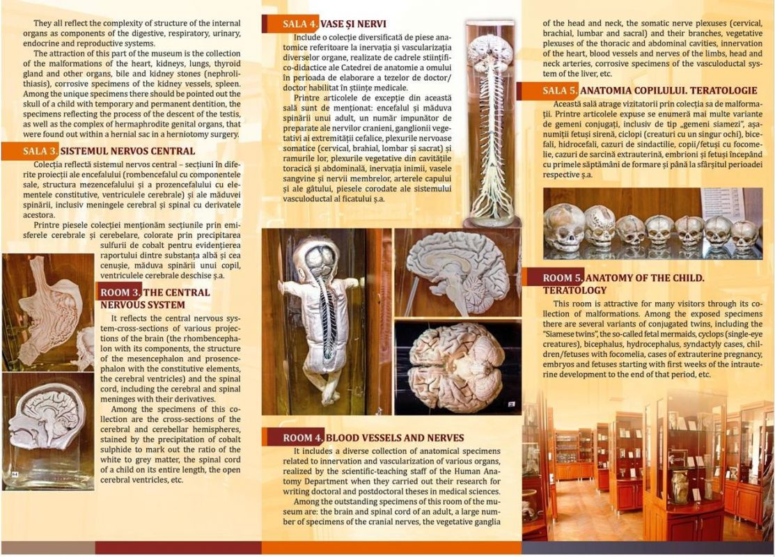
Room 5. Anatomy of the child
Of all the museum rooms, the latter seems to attract the most visitors and amaze them with an impressive collection of malformations.
The exhibited pieces include several variants of conjugated twins, including the type "Siamese twins", such as mermaid fetuses, cyclops (one-eyed creatures), bicephalous, hydrocephalus, syndactyly cases, children with focomelia, cases of extrauterine pregnancy. , embryos and fetuses from the first weeks of training to the end of the respective period, etc.
With images of impeccable quality of the anatomical pieces made by the scientific-didactic staff of the Department (university professors Boris Z. Perlin, Vasile N. Andrieş, Natalia V. Cherdivarenco, Victor T. Jița, Mihail I. Ştefaneţ, Ilia M. Catereniuc, university lecturers Galina V. Vincenco, Tatiana A. Iastrebova, Alexei V. Popa, Tamara M. Titov, Emilia V. Gherghelegiu-Poburnaia, Teodor I. Lupaşcu, Eugenia S. Lopotencu, Olga V. Belic, Dumitru Gh. Batîr, Anatolie N. Nastas, university assistants Ilia A. Gurițenco, Domnica M. Stratilă, etc.), over the years are illustrated many specialized editions, published in the country and abroad, including the famous atlases of Human Anatomy, edited by R. Д. Sinelnikov (Синельников Р. Д., Синельников Я. Р. Атлас анатомии человека. Москва: Медицина, any ed.) And the one of the vegetative nervous system, under the editorship of PI Lobko (Л П Побер) Денисов С. Д., Пивченко П. Г. Вегетативная нервная система. Атлас. Mинск, 1988), monographs, textbooks and other methodological-didactic works, elaborated at the cathedra.